


This year’s Nobel Prize in Physiology or Medicine went to Karl Deisseroth, Peter Hegemann and Georg Nagel for their work on optogenetics: the ability to control the activity of cells with light. The technique uses light-sensitive proteins to switch specific cells on or off, mainly neurons, and belongs to the world of research.
In the skin, we don’t practise optogenetics in the strict sense, but the underlying principle is a very familiar one: cells detect light, and that light can change what they do. In dermatology, we have been making use of it for decades.
Skin responds to light too
Cells don’t have eyes, but they do have molecules able to absorb certain wavelengths, and each type of cell reacts in its own way. The fibroblast, the cell that produces collagen, is a good example. As we age we lose collagen, and the skin thins until it becomes atrophic, like paper. In one laboratory experiment, fibroblasts were grown in several dishes and each received a different colour of light: yellow, green, blue, ultraviolet and red. Under ultraviolet light they gradually declined, which helps explain why the sun wears these cells down and why we have more wrinkles in the areas most exposed to it. They grew best under red light.
The explanation lies in the mitochondria, the engines of the cell, which burn oxygen to produce energy. They contain enzymes with copper atoms that absorb red light and, in doing so, increase their activity, a little like letting more oxygen into an engine. This has been known for a long time: in 1967, an experiment in mice was published in which hair grew faster in the areas exposed to red light, and we know that wounds heal more quickly with it.
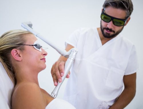
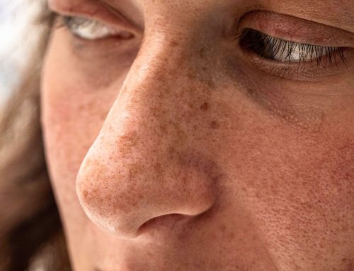
But the fibroblast is only one of the skin cells that responds to light. The melanin in melanocytes and pigmented keratinocytes absorbs the light of specific lasers, which break the pigment up in certain kinds of pigmentation. The haemoglobin in red blood cells absorbs the light of vascular lasers and intense pulsed light, which makes it possible to treat the redness of rosacea and dilated vessels. Melanin in the hair carries heat to the follicle in laser hair removal. Water in the tissue absorbs the light of fractional CO2 laser, which creates microscopic columns of controlled injury: the epidermis regenerates from the intact skin and fibroblasts produce new collagen, which is key in acne scarring. And in acne, the bacterium Cutibacterium acnes produces porphyrins, molecules that absorb blue light, which is why certain light sources can help reduce the bacteria and the inflammation.
Two ways of using light
When light reaches the skin, what happens depends on three variables: the wavelength, the irradiance and the exposure time. Their combination gives rise to the two effects we use in dermatology.
The first is the photobiological effect, or photobiomodulation. It is produced with low-irradiance light applied over several minutes, a dose low enough not to heat the tissue. What distinguishes it is that each wavelength is absorbed by different molecules in the cell, such as cytochrome c oxidase in the mitochondria, opsins, flavins, cryptochromes or porphyrins. When they absorb the light, these molecules set different pathways in motion, with changes in ATP production, reactive oxygen species and the release of nitric oxide, which ultimately regulate cell proliferation, migration and collagen synthesis. That is why different types of light give us different results, and why the depth the light reaches and the dose required (measured in J/cm²) also change: below a certain threshold there is no effect, and above it the benefit can be lost. It is used to help wounds repair, stimulate skin growth and rejuvenate the skin. Many of these mechanisms are still not fully confirmed, and much of the evidence comes from in vitro and animal studies.
The second is the photothermal effect, or selective photothermolysis. Here the light is delivered in very short, high-energy pulses, and the principle requires three things: a wavelength absorbed by the structure we want to treat (melanin, haemoglobin or water), a pulse duration shorter than that structure’s thermal relaxation time, so the heat stays confined to it, and sufficient energy. The target heats up while the surrounding tissue is spared. The aim is not to modulate the function of the cell but to injure a specific structure in a controlled way, and the repair response follows from that.
Which light does your skin need?
At De Felipe Dermatology Clinic, we have worked with different light sources and lasers for decades. That experience is most evident in our holistic approach to the different conditions of the skin, where we combine light sources with other therapies to achieve optimal results that last over time.
Light is a tool of great precision, but every concern and every skin responds differently. That is why we always begin with an individualised evaluation, which determines the best treatment plan for your skin.